When a dog is limping but X-rays do not explain the pain, or a cat has weight loss and abnormal bloodwork, advanced imaging can provide answers that a physical exam alone cannot. In the pet CT scan vs ultrasound decision, the best test is not necessarily the newest or most detailed one. It is the test that can most clearly answer your veterinarian’s specific medical question while keeping your pet’s comfort, safety, and needs in focus.
At AV Veterinary Center, advanced diagnostics are part of a personalized care plan. Our team considers your pet’s symptoms, exam findings, prior test results, age, and overall health before recommending imaging. That approach helps avoid unnecessary procedures while moving efficiently toward a diagnosis and treatment plan.
What an Ultrasound Shows
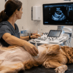
Ultrasound uses sound waves to create real-time images of internal organs and soft tissues. A veterinarian can watch structures move as the scan is performed, making ultrasound especially useful for evaluating the abdomen, chest, heart, and certain soft tissue areas.
For example, an abdominal ultrasound may help assess the liver, spleen, kidneys, bladder, intestines, pancreas, lymph nodes, and reproductive organs. It can identify changes such as enlarged organs, fluid in the abdomen, some masses, stones, cysts, inflammation, or signs of obstruction. If a suspicious area is found, ultrasound may also help guide a needle sample or biopsy with greater precision.
Ultrasound is often a strong first choice when a pet has vomiting, diarrhea, poor appetite, abdominal discomfort, unexplained weight loss, changes in urination, or abnormal bloodwork. Cardiac ultrasound, called echocardiography, is used to evaluate the heart’s structure and function when a murmur, abnormal rhythm, coughing, or other concern is present.
A major advantage is that ultrasound does not use radiation. Many pets can tolerate a routine ultrasound while awake with gentle handling, although sedation may be recommended for anxious, painful, or uncooperative patients. The fur over the area being examined is usually clipped so the ultrasound probe can make proper contact with the skin.
Where Ultrasound Has Limits
Sound waves do not travel well through air or dense bone. That means ultrasound is less effective for seeing through the lungs, inside the skull, or through parts of the body obscured by intestinal gas. It also provides a smaller field of view than CT and depends heavily on the skill of the person performing and interpreting the scan.
Ultrasound can reveal that a mass is present, but it may not always show its full extent or whether it has spread to nearby structures. In those cases, CT may provide the more complete roadmap.
What a CT Scan Shows
Computed tomography, or CT, uses a series of X-ray images and computer processing to create detailed cross-sectional views of the body. Think of it as looking at the body in thin slices rather than as a single flat image. A CT scan can also create three-dimensional images that are valuable for surgical planning.
CT is particularly helpful for evaluating bone, the skull, nasal passages, ears, teeth, spine, chest, joints, and complex soft tissue structures. It is often used when conventional X-rays show an abnormality but cannot define it fully. For a pet with chronic nasal discharge, for instance, CT can help evaluate nasal disease, infection, foreign material, tooth-root problems, or a possible tumor.
It can also be a vital tool in trauma care. CT may help identify subtle fractures, assess the extent of injuries, and give the surgical team a clearer view before orthopedic, neurologic, or soft tissue procedures. For pets with suspected tumors, a contrast-enhanced CT can help determine the size and location of a mass, its relationship to nearby tissues, and whether there are signs of spread to the lungs or lymph nodes.
Because CT is fast, it can be an excellent option for patients who need detailed imaging promptly. The scan itself may take only minutes. However, pets need to remain completely still for accurate images, so general anesthesia is commonly required. This allows the veterinary team to position the patient safely and obtain clear diagnostic images without causing stress or discomfort.
Why Contrast May Be Used
Some CT examinations include an injectable contrast agent that makes blood vessels, organs, inflammation, and certain masses easier to distinguish. Not every patient needs contrast, and it is not appropriate in every situation. Your veterinarian will review your pet’s medical history, kidney function, hydration status, and the purpose of the scan before making that recommendation.
Pet CT Scan vs Ultrasound: Key Differences
The most practical difference is the type of detail each study provides. Ultrasound excels at evaluating many abdominal organs in real time and can guide sample collection. CT offers a broader, highly detailed view of complex anatomy, especially bone, air-filled spaces, and areas that are difficult to assess with ultrasound.
Neither test replaces the other. A pet with increased liver enzymes may benefit first from ultrasound because it can closely assess the liver, gallbladder, and surrounding abdominal organs. A pet with a complicated fracture, spinal concern, middle-ear disease, or nasal mass may be better served by CT because it can show the anatomy from multiple angles and support treatment planning.
The need for anesthesia is another meaningful distinction. An ultrasound may be completed with minimal restraint, while CT generally requires anesthesia. Anesthesia always deserves thoughtful consideration, particularly for senior pets or those with heart, lung, kidney, or liver disease. Before anesthesia, a veterinarian may recommend bloodwork, chest imaging, or other screening based on the individual patient.
Cost can differ as well. CT requires specialized equipment, anesthesia, and advanced image interpretation, so it is usually more expensive than a standard ultrasound. Still, choosing the imaging test most likely to answer the question can prevent delays, repeat procedures, and uncertainty. The goal is not to order the most advanced test by default. It is to select the test that gives your pet the right information at the right time.
How Veterinarians Choose the Right Imaging Test
Your veterinarian starts with the problem, not the machine. A complete history and physical examination help narrow the possibilities. Bloodwork, urinalysis, X-rays, and other initial diagnostics may point toward a specific organ system or condition.
If the concern involves abdominal organs, fluid, pregnancy, or a soft tissue structure near the surface, ultrasound may be recommended first. If the concern involves the braincase, nasal cavity, bones, lungs, spine, complex fractures, or surgical mapping, CT may be more appropriate. In some cases, both tests are useful because they answer different questions.
For example, a dog with a suspected abdominal mass may have an ultrasound to identify the affected organ and obtain a sample. CT may follow if surgery is being considered and the team needs to understand the mass’s full size, blood supply, or possible spread. Similarly, a cat with chronic ear disease may need CT to evaluate the middle ear and surrounding bone before a specialized ear procedure.
Preparing Your Dog or Cat for Imaging
Preparation depends on the planned study and whether sedation or anesthesia is needed. Your veterinary team will give you specific instructions, including whether your pet should fast before the appointment. Fasting is commonly required for anesthesia and may also improve the quality of abdominal imaging by reducing food in the stomach.
Bring a list of current medications, supplements, recent medical records, and any previous imaging results if they were completed elsewhere. Let the team know about known allergies, prior reactions to sedation or anesthesia, chronic health conditions, and changes you have noticed at home. Details such as when symptoms began, whether they are getting worse, and what triggers them can be just as helpful as the images themselves.
If your pet is painful, having trouble breathing, collapses, develops a swollen abdomen, cannot urinate, or has severe vomiting or diarrhea, seek veterinary care promptly rather than waiting for a routine imaging appointment. Advanced diagnostics can be valuable, but stabilizing an emergency patient always comes first.
Questions Worth Asking Before Imaging
Clear communication can make an unfamiliar recommendation feel more manageable. Ask what question the scan is intended to answer, whether another test could provide the same information, and how the results may change your pet’s treatment. It is also reasonable to ask whether sedation or anesthesia is required, what safety screening is recommended, and when results are expected.
A good imaging recommendation should connect directly to a next step. That may mean confirming a diagnosis, collecting a sample, planning surgery, monitoring a known condition, or determining that treatment can remain conservative.
When your pet needs more than a routine exam can reveal, you deserve a team that explains the options with care and clinical clarity. A thoughtful conversation with your veterinarian can help you choose an imaging plan that protects your pet’s comfort while bringing your family closer to the answers they need.